
An electrocardiogram (ECG) is a test that can help diagnose certain heart conditions by measuring the electrical activity of the heart. An ECG can help diagnose certain heart conditions, including abnormal heart rhythms and coronary heart disease (heart attack and angina). A doctor may recommend an ECG if you are experiencing symptoms like chest pain, breathlessness, dizziness, fainting or a feeling of your heart racing, fluttering, thumping or pounding in your chest (palpitations). An ECG can also help monitor how treatments for a heart condition, like medicines or implantable cardiac devices, are working. Resting ECG – you lie down for this type of ECG. No movement is allowed during the test, as electrical impulses from other muscles can interfere with the test. This type of ECG usually takes five to 10 minutes. An ECG can help diagnose: • conditions involving the heart’s electrical system •heart attacks •abnormal heart rhythms (arrhythmia) – rapid, slow or irregular heart beats •poor blood supply to the heart •heart inflammation (pericarditis or myocarditis) •cardiac arrest
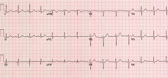
A normal ECG is illustrated above. Note that the heart is beating in a regular sinus rhythm between 60 - 100 beats per minute (specifically 82 bpm). All the important intervals on this recording are within normal ranges. 1. P wave: upright in leads I, aVF and V3 - V6 normal duration of less than or equal to 0.11 seconds polarity is positive in leads I, II, aVF and V4 - V6; diphasic in leads V1 and V3; negative in aVR shape is generally smooth, not notched or peaked . 2. PR interval: Normally between 0.12 and 0.20 seconds. 3. QRS complex: Duration less than or equal to 0.12 seconds, amplitude greater than 0.5 mV in at least one standard lead, and greater than 1.0 mV in at least one precordial lead. Upper limit of normal amplitude is 2.5 - 3.0 mV. small septal Q waves in I, aVL, V5 and V6 (duration less than or equal to 0.04 seconds; amplitude less than 1/3 of the amplitude of the R wave in the same lead). represented by a positive deflection with a large, upright R in leads I, II, V4 - V6 and a negative deflection with a large, deep S in aVR, V1 and V2 in general, proceeding from V1 to V6, the R waves get taller while the S waves get smaller. At V3 or V4, these waves are usually equal. This is called the transitional zone. 4. ST segment: isoelectric, slanting upwards to the T wave in the normal ECG can be slightly elevated (up to 2.0 mm in some precordial leads) never normally depressed greater than 0.5 mm in any lead 5. T wave: T wave deflection should be in the same direction as the QRS complex in at least 5 of the 6 limb leads normally rounded and asymmetrical, with a more gradual ascent than descent should be upright in leads V2 - V6, inverted in aVR amplitude of at least 0.2 mV in leads V3 and V4 and at least 0.1 mV in leads V5 and V6 isolated T wave inversion in an asymptomatic adult is generally a normal variant 6. QT interval: Durations normally less than or equal to 0.40 seconds for males and 0.44 seconds for females.
The ST segment on an electrocardiogram (ECG) normally represents an electrically neutral area of the complex between ventricular depolarization (QRS complex) and repolarization (T wave). However, it can take on various waveform morphologies that may indicate benign or clinically significant injury or insult to the myocardium. Understanding the differential diagnosis for variations in the ST segment is critical for clinical management as it can influence treatment. This article summarizes ST segment, including how it is defined, measured, and how it varies. This article also examines and summarizes ST-segment morphologies unique to various conditions that present with ST elevation or depression.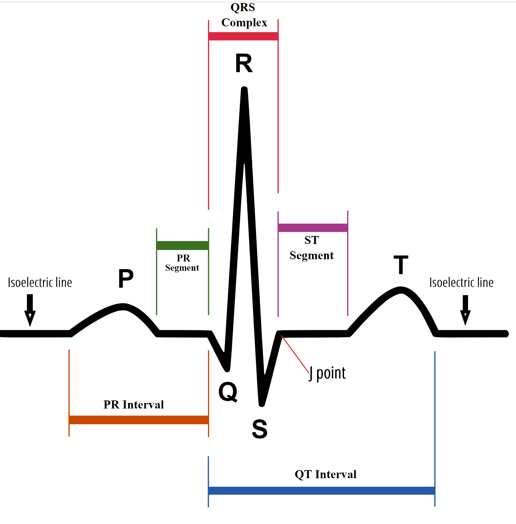
The procedure for taking an exercise stress test is relatively straightforward. As you walk on a treadmill at progressively faster speeds, an electro-cardiograph measures your heart rate and the electrical activity in your heart, and your blood pressure is recorded periodically. 0 = Upsloping 1 = flat 2 = downsloping
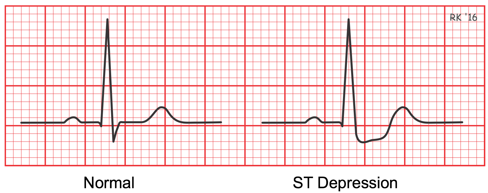
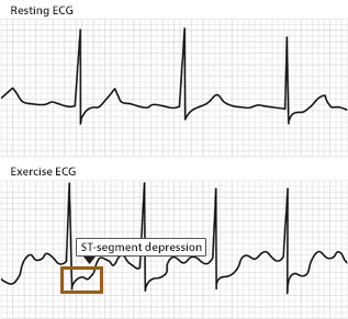
The treadmill electrocardiogram (ECG) stress test is widely used to screen for obstructive coronary artery disease (CAD). The presence of STsegment changes, either depression or elevation, on the ECG during the treadmill test often suggests presence of CAD and warrants further management. We herein present three cases, with evidence of ischaemia on the treadmill ECG stress test. In addition, we discuss the use of the treadmill ECG stress test, including its indications, contraindications, reasons for termination and interpretation of the ST-segment changes, heart rate, as well as blood pressure responses to exercise.
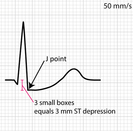
Duration of the segment is very important because it needs to be checked that after peak stress, the recovery is happening constantly or not with a positive treadmill test. The abnormal values come under the downslope of depression with less than or equal to 1 mm with 60 to 80 ms.\n\n 0= Normal 1= fixed defect 2= reversible defect 3= defect
This test, which involves a small amount of a radioactive substance injected intravenously, measures the blood flow to your heart when you’re resting and active. A thallium stress test is an imaging test that indicates how well blood flows into your heart while you’re exercising or at rest. It’s also called a nuclear stress test. During the procedure, a small amount of thallium, a radioactive tracer, is administered into a vein on your arm. The tracer is a dye that makes your blood flow visible to a special camera called a gamma camera. This camera can reveal any issues your heart muscle may be having. Flat metal disks called electrodes are placed on your chest, arms, and legs and connected to an electrocardiogram (EKG) machine that records your heart’s electrical activity. A doctor may order a thallium test for a variety of reasons, including: • if they suspect your heart isn’t getting enough blood flow when it’s under stress — for example, when you exercise • if you have chest pain or worsening angina • if you’ve had a heart attack • to check how well medications are working • to determine whether a procedure or surgery was successful • to determine whether your heart is healthy enough to start an exercise program The thallium stress test can show: • the size of your heart chambers • how effectively your heart pumps (ventricular function) • how well your coronary arteries supply your heart with blood, known as myocardial perfusion • if your heart muscle is damaged or scarred from previous heart attacks A thallium stress test is different from an exercise stress test, which only uses electrodes connected to an EKG to record your heart’s electrical activity as you exercise. A thallium stress test is also different from a stress echocardiography, during which electrodes are connected to an electrocardiograph (ECG) that uses ultrasound to provide images of your heart as you exercise. How is a thallium stress test performed? A thallium stress test must be done at a hospital, medical center, or doctor’s office. The test usually takes about 3 to 4 hours. A nurse or healthcare professional inserts an intravenous (IV) line, usually on the inside of your elbow. Thallium is injected through the IV. This radioactive tracer dyes your blood flow so it can be detected by the gamma camera. The test includes exercise and resting portions, and your heart is photographed during both. The doctor administering your test will determine the order in which these tests are performed. You’ll receive an injection of thallium before each portion. Resting portion During this part of the test, you lie down for 15 to 45 minutes while the radioactive tracer works its way through your bloodstream to your heart. You then lie down on an exam table with your arms above your head, and a gamma camera above you takes pictures. Exercise portion In the exercise portion of the test, electrodes connected to an EKG machine are applied to your chest, arms, and legs. You then walk on a treadmill or pedal an exercise bicycle. Most likely, the doctor will ask you to start slowly and progressively pick up the pace into a jog. You may need to run on an incline to make it more challenging. If you’re unable to exercise, the doctor will give you a medication that stimulates your heart and makes it beat faster. This simulates how your heart would act during exercise. The gamma camera records pictures that show the flow of blood through your heart. Your blood pressure and heart rhythm are monitored while you exercise. Once your heart is working as hard as it can, you’ll receive another thallium injection and wait 15 to 40 minutes for your heart to absorb the tracer. You’ll then resume exercising. The doctor will compare these pictures with the set of resting images to evaluate how weak or strong the blood flow to your heart is.